ABSTRACT: 1227
Brain Activation and Dental Wear Correlations in Bruxers and Non-Bruxers
| K.E. BYRD1, M. DZEMIDZIC1, L.M. ROMITO2, T.M. TALAVAGE3, and D. WONG1, 1Indiana University School of Medicine, Indianapolis, USA, 2Indiana University School of Dentistry, Indianapolis, USA, 3Purdue University, West Lafayette, IN, USA |
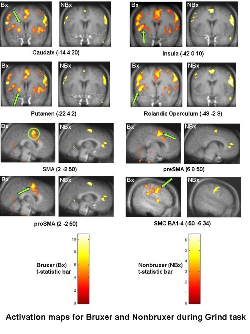
Objectives: To compare the brain activation patterns evoked in bruxers (Bx) and nonbruxers (NBx) while executing brux-like tasks during fMRI scanning. Methods: Twenty subjects (5M, 5F per group; mean ±sd age of 26.0 ± 4.0 years) received an oral exam and dental impressions for evaluation of their dental wear. A custom oral appliance was affixed to maxillary anterior teeth to minimize head motion but permitted interference-free mandibular motion during imaging. Subjects performed two oromotor tasks of clench (C) and grind (G) in a block-design paradigm while being imaged in a 3T Siemens scanner. Task and group-dependent effects were evaluated using SPM5 software. Results: A neural network consisting of cortical and subcortical motor regions was observed in both groups for the two oromotor tasks. A region of interest analysis of mean percent signal change revealed significant differences (p<0.05, p<0.01) between groups in six a priori defined regions (caudate, insula, putamen, rolandic operculum, supplementary motor area, and sensorimotor cortex). Mean percent signal changes regressed against degree of dental wear within and between groups also showed significant differences (p<0.05, p<0.01). Conclusion: A functional neuroimaging approach was used to elucidate the brain regions activated in bruxer and matched control subjects performing oromotor movements that mimic bruxism. Group comparison of brain activity in specific regions may provide insights into the dynamics of bruxism and its clinical manifestations such as dental wear. These analyses of brain activity correlated with dental wear also provide new understanding of possible sensorimotor feedback or affective components underlying bruxism. In addition, these techniques can be extended to functional brain imaging of TMJMD subjects and associated comorbidities. Funding provided by American Equilibration Society (AES # 2005-2) and pilot grants from the Departments of Radiology, Anatomy & Cell Biology, and the Oral Health Institute, Indiana University Schools of Medicine and Dentistry.
|
| Seq #174 - Poster Presentations 10:45 AM-12:00 PM, Saturday, April 5, 2008 Hilton Anatole Hotel Trinity I - Exhibit Hall |
©Copyright 2008 American Association for Dental Research. All Rights Reserved.