ABSTRACT: 0434
Effects of Age and Exercise on Bone Tissue Quality
| S.R. OBREITER, N.D. SAHAR, M.S. RON, L.A. WINKEL, and D.H. KOHN, University of Michigan, Ann Arbor, USA |
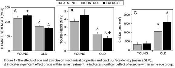
Objective: This study analyzed the effects of age and exercise on microdamage and tissue level mechanical properties in bone. Methods: Young and old (4 and 18 months) C57BL/6 male mice were divided into two weight-matched groups of control and exercise (run on a treadmill 30 min/day, 12m/min, 5° incline). After 3 weeks, a randomly selected tibia from each mouse was harvested and scanned using micro-computed tomography to determine geometric properties and bone mineral density. Half of the tibiae were mechanically tested under monotonic four-point bending while monitoring force and displacement allowing measurement of strength, toughness, ductility, and modulus. Remaining tibiae were stained with basic fuchsin, imbedded in PMMA, and sectioned transversely from the mid-diaphysis (width of 120–150µm). Laser scanning confocal microscopy images were analyzed for two types of microdamage: microcracks (quantified by number and length) and diffuse damage (quantified by area). Effects of age and exercise were determined using a 2-way ANOVA, with a Student-Newman-Keuls post hoc test and significance set at p<0.05. Results: Exercise increased tissue strength (p=0.042) in young mice, but had no significant benefit in old bones, and deceased toughness. Aging resulted in significantly reduced strength (p≤0.001), toughness (p≤0.001), and ductility (p=0.022); and also resulted in increased total crack length (p=0.018).
Conclusions: This exercise model was beneficial to skeletal integrity in young mice, but provided no significant gain in old mice. Mechanical integrity was decreased in old animals. Mechanical properties were taken at the tissue level indicating that skeletal integrity in old mice was compromised by degradation of quality and not loss of bone. Acknowledgements: DoD/US Army DAMD17-030100556 and University of Michigan |
| Seq #69 - Structure/Function of Protein and Mineral 2:45 PM-3:45 PM, Thursday, April 3, 2008 Hilton Anatole Hotel Trinity I - Exhibit Hall |
©Copyright 2008 American Association for Dental Research. All Rights Reserved.