ABSTRACT: 0641
Comparison of histology techniques used for histomorphometry of cranial defects
| N. MEHTA, and L.A. OPPERMAN, Texas A&M Health Science Center, Baylor College of Dentistry, Dallas, USA |
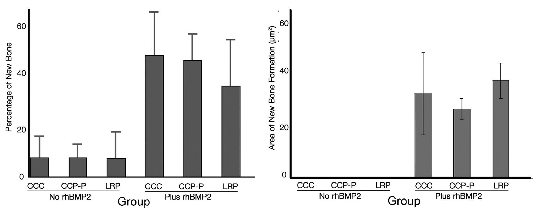
Objectives: In order to protect the brain during the regeneration of a bony cranial defect, several combinations of materials are used to spur rapid regeneration of bone. A previous study used undecalcified sections to assess amounts of new bone formation in large cranial defects. This study used histomorphometry of paraffin sections to examine the quantity of new bone formation compared to methylmethacrylate embedded sections from the same animals. Methods: A substantial portion of the cranial vault of eighteen dogs was removed, followed by immediate replacement with demineralized bone matrix. Nine dogs were supplemented with recombinant human BMP2 (rhBMP2), while the other nine did not receive rhBMP2. In each group, three cranial vaults were fixed with Lactosorb® resorabable mesh (LRM), and six cranial vaults were fixed with cobalt chrome plates (CCP), with addition of platelet-rich plasma in three of the latter (CCP-P). Necropsy was conducted at 12 weeks and crania dissected and prepared for H&E sectioning and staining. Histomorphometry was used to calculate the amount of new bone growth. Results: With both histology techniques, the group with no rhBMP2 had minimal measurable new bone within the defects. In undecalcified specimens CCP-rhBMP-2 gave the highest of amount of new bone regeneration, followed by CCP-P-rhBMP2, and LRM-rhBMP2 the least amount of new bone (Figure on left). In the paraffin sections, LRM-rhBMP2 specimens showed greater bone area than CCP-rhBMP2 or CCP-P-rhBMP2 (Figure on right). However, there were no significant differences between the groups, and no significant differences in scoring between the methods of tissue preparation. Conclusions: When tissue samples are so small that multiple processing methods cannot be done, either decalcified or undecalcified histology alone is sufficient for assessing amounts of new bone formation. Supported by a grant from Medtronic.
|
| Seq #105 - DENTSPLY/Caulk - Clinical Science Category 1:30 PM-2:30 PM, Friday, April 4, 2008 Hilton Anatole Hotel Trinity I - Exhibit Hall |
©Copyright 2008 American Association for Dental Research. All Rights Reserved.