ABSTRACT: 2236
Correlations between Cephalometric and Photographic Measurements of Facial Attractiveness
| H.S. OH, University of the Pacific, Arthur A. Dugoni School of Dentistry, San Francisco, CA, USA, T.-M. XU, Peking University School of Stomatology, Beijing, China, H. PEARSON, New Jersey Dental School - UMD, Newark, USA, and S. BAUMRIND, University of the Pacific, San Francisco, CA, USA | |
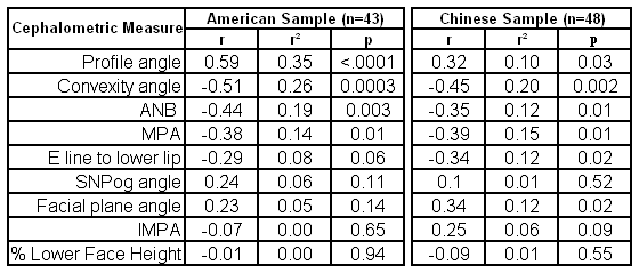
Objective: To investigate the extent to which selected cephalometric measurements correlate with rankings of facial attractiveness on end-of- treatment facial photographs of Chinese and white US orthodontic patients. Method: Similar samples of standardized end-of-treatment facial photographs of Chinese and white US orthodontic patients were drawn randomly at the Departments of Orthodontics at Peking University and the New Jersey Dental School, Newark. Eight groups of 12 patients each were ranked by 20 U.S. and 25 Chinese orthodontists. Each judge assigned a rank to each patient in each group, ranging from 12 for “most attractive” to 1 for “least attractive”. Mean ranks for each patient were calculated separately for the Chinese and U.S. judges. End-of-treatment lateral cephalograms taken at the same time as the photographs were “traced” independently by multiple judges for both American and Chinese samples. Nine cephalometric measures usually considered to reflect facial attractiveness were correlated with the photographic rankings of facial attractiveness separately for each ethnicity. Results: For the American sample, the strongest correlation was found in soft tissue profile angle (r=0.60, r2=0.35, p<0.0001). The next strongest variables were convexity angle (r=0.-52, r2=0.26, p=0.0003), and ANB angle (r=-0.44, r2=0.19, p=0.003). For the Chinese sample, the strongest correlation was found in convexity angle (r= - 0.45, r2 =0.20, p=0.002).
Conclusion: In general, correlations between cephalometric variables and rankings of facial attractiveness were less strong than had been expected. Soft tissue cephalometric measures performed slightly better than hard tissue measures but no cephalometric measure accounted for more than 1/3 of the variance in the photographic rankings. | |
| Seq #214 - 2D and 3D Craniofacial Imaging and Analyses 2:00 PM-3:15 PM, Friday, July 4, 2008 Metro Toronto Convention Centre Exhibit Hall D-E | |
©Copyright 2008 American Association for Dental Research. All Rights Reserved.
