ABSTRACT: 0172
Biochemical identification of pre-malignant changes in oral mucosal lesions
| M.J. GERMAN1, C.M. ROBINSON1, J.M. NICHOLSON2, and P.J. THOMSON1, 1Newcastle University, Newcastle upon Tyne, United Kingdom, 2Science & Technology Facilities Council, Warrington, United Kingdom | |
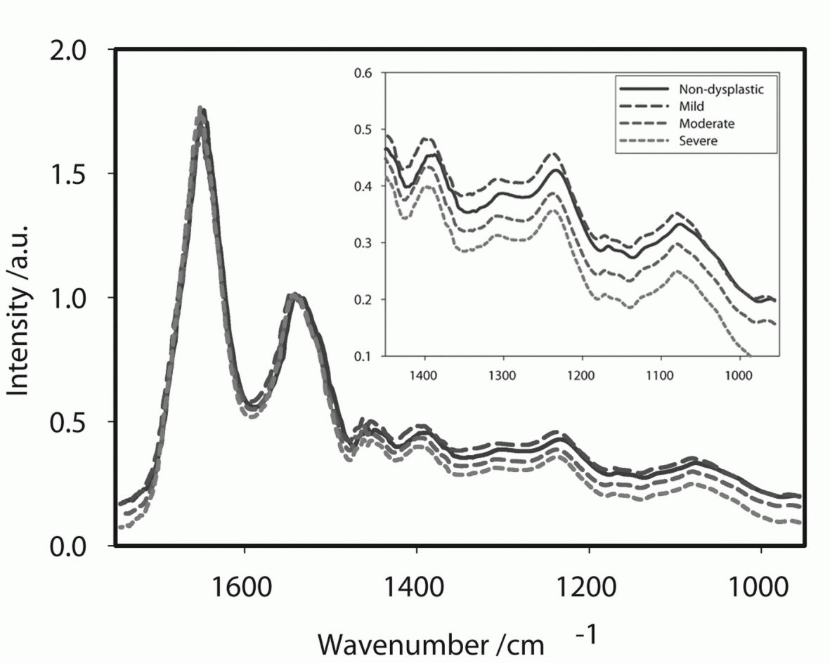
Click on images to view full size. Objectives: To establish whether infra-red (IR) microscopy could be used as a diagnostic tool by looking for biochemical markers indicative of change in state from normal to dysplasia in oral mucosal biopsies. Methods: Oral mucosal biopsies were categorised into four groups, namely, non-dysplastic, mild, moderate, and severe dysplasia. Five biopsies per group were selected and 10 mm thick sections were cut and mounted on BaF2 slides for IR analysis. Areas for analysis were identified using parallel H & E stained sections and spectra were taken from the basal epithelium for each specimen. Using an IR microscope interfaced to a synchrotron source (Daresbury, UK) spectra were taken from 10 x 10 mm area in transmission mode from 25 individual cells for each specimen. Results: We were able to show that there are differences between the spectral responses of basal keratinocytes in specimens from each of the categories; the mean spectra for each group are summarised in Fig 1. With increasing epithelial dysplasia there was a reduction in the intensity of peaks related to the concentrations of RNA (1121 cm-1), DNA (1020 cm-1), carbohydrates (900-1185 cm-1) and nucleic acid phosphates (1185-1300 cm-1). One-way ANOVA revealed that there were no statistically significant differences in intensities of these peaks between the non-dysplastic and mild dysplasia samples. There were significant differences between all of the other samples and the moderate and severe dysplasia samples for the RNA, DNA and carbohydrate peaks (p<0.05). Conclusion: We have shown a correlation between the biochemical properties of oral keratinocytes and conventional histological grading of dysplastic lesions. This suggests that IR spectroscopy may be a useful tool in the objective assessment of oral dysplastic lesions. Figure 1: Mean spectra for the four groups
| |
| Seq #39 - Head and Neck Cancer, Dental Implants 9:00 AM-10:30 AM, Thursday, July 3, 2008 Metro Toronto Convention Centre Room 709 | |
©Copyright 2008 American Association for Dental Research. All Rights Reserved.