ABSTRACT: 0968
In Vitro Study of Polypeptide Catalyzed Biosilicification of Dentin Surfaces
| R.B. KAZEMI1, M.C. ADVINCULA1, T. KOMABAYASHI1, P. PATEL2, P.T. MATHER3, D.G. GOBERMAN4, and A.J. GOLDBERG1, 1University of Connecticut Health Center, Farmington, USA, 2Case Western Reserve University, Cleveland, OH, USA, 3Syracuse University, NY, USA, 4University of Connecticut, Storrs, USA | |||||||||||||||||||||||||
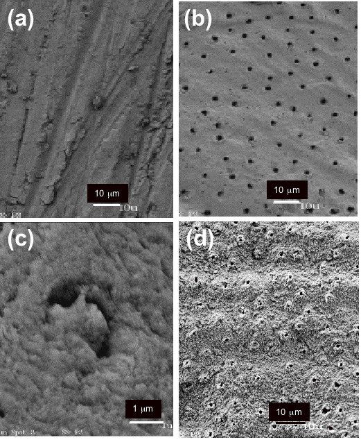
Introduction: Resin bonding agents may deteriorate with time and become ineffective in sealing restorative-tooth interfaces and in preventing dentin hypersensitivity. In situ formation of mineral particles to occlude dentin tubules might reduce such clinical failures. As a model system we investigated formation of silica on dentin from tetramethylorthosilicate (TMOS) by the amine-containing macromolecular catalysts polylysine (PLL), an analog of the protein catalyst responsible for silica formation in primitive marine species. Objectives: To evaluate if biocatalyzed silicification could be conducted on a dentin surface and occlude dentine tubules. Methods: Dentin disks (n=6) nominally 500Ám thick were prepared from defect/caries-free human molars. After removal of smear layer 1mM 30-70kDa PLL(Aldrich) was applied layer-by-layer with HCl pre-hydrolyzed 100mM TMOS. Smear-free disks(n=4) served as controls. Disks were examined with SEM (JSM-6320F,JEOL) and XPS (VG Escalab MkII). The permeability of four smear-free disks was measured at baseline and after silicification, and results were compared with a matched-pair t-test. Results: Smear (Fig-a) and smear-free (Fig-b) morphologies were as expected. The PLL/TMOS treated disks exhibited a partial coating of 0.2Ám particles (Fig-c), whose morphology and XPS atomic percentages (below) were consistent with silica.
Mean(sd) permeability decreased from 1.40(2.40) to 0.56(0.97)x10-3 ÁL/cm2-min-cmH2O, but the difference was not statistically significant (p=0.325), possibly due to technique, dentin variation, insufficient mechanical integrity or adhesion of the particles, which were pushed from the tubules (Fig-d). Conclusions: The results demonstrate that the polypeptide retained its catalytic activity on dentin and the formed particles bridged the patent tubules. Further understanding of the reaction parameters for silica or other biocatalyzed minerals could provide control of particle properties and location of deposition for novel sealing or resin reinforcement applications.
| |||||||||||||||||||||||||
| Seq #106 - Dentin-Bonding Interface 3:30 PM-4:45 PM, Thursday, July 3, 2008 Metro Toronto Convention Centre Exhibit Hall D-E | |||||||||||||||||||||||||
|
Back to the Dental Materials 2: Adhesion - Leakage/Margin Assessments Program | |||||||||||||||||||||||||
©Copyright 2008 American Association for Dental Research. All Rights Reserved.
